Back Of Skull Anatomy : Skull Notes. Hank grebe / getty images See anatomy of the head and neck stock video clips. At the base of the skull is bone that supports 4 brain components—the frontal lobe, temporal lobe, brain stem, and cerebellum. Anatomy the base of the skull is a complex area. 1 also, some people are more prone to headaches than others.
ads/bitcoin1.txt
The skull is a strong, bony capsule that rests on the neck and encloses the brain. The brain is connected with other anatomical structures by the nerves and blood vessels going through many foramina, and the largest foramen of the skull called the foramen magnum. It is located next to five of the cranium bones. The upper jaw, but not the lower, is part of the skull. This portion of the skull base consists of the orbital portion of the frontal bone.
Human Head Skull And Cervical Vertebrae Rear View Stock Photo Download Image Now Istock from media.istockphoto.com Front anatomical view of human skull bone with the vault of the skull seperated by saw and without mandible in isolated white back. Muscle head anatomy vocal organ diagram female neck anatomy neck wireframe head neck human anatomy head artery anatomy face pharynx vector neck degree head anatomy 3d. It is trapezoidal in shape and curved on itself like a shallow dish. The mayo clinic says that these factors can be chemical activity in your brain, the nerves of blood vessels surrounding your skull, or the muscles at the back of the head and neck. Skull, skeletal framework of the head of vertebrates, composed of bones or cartilage, which form a unit that protects the brain and some sense organs. Cranium) is the skeleton of the head composed of 22 separate bones joined together primarily by sutures. The occipital bone is the only bone in your head that connects with your cervical spine (neck). It is made up of more than 100 billion nerves that communicate in trillions of connections called synapses.
Encephaloceles can occur in the base of the skull, the top or back of the skull, or between the forehead and nose.
ads/bitcoin2.txt
The skull base offers support from the bottom of the brain. The pain in the back of your head occurs when one or a combination of factors affect your brain. The 22nd bone is the mandible (lower jaw), which is the only moveable bone of the skull. It also covers some common conditions and injuries that can affect the back. 1 also, some people are more prone to headaches than others. Numerous muscles, ligaments and tendons support the spine, providing it with flexibility and a great range of motion. The skull is a strong, bony capsule that rests on the neck and encloses the brain. Hank grebe / getty images Bone back of head bigger on one side, bone back of the head, bone on back of skull, bone spur back of skull, skull bone back of head, bone, bone back of head bigger. Conditions associated with encephaloceles include hydrocephalus (excess accumulation of cerebrospinal fluid in the brain), developmental delays, microcephaly (an abnormally small head), paralysis and seizures. Muscle head anatomy vocal organ diagram female neck anatomy neck wireframe head neck human anatomy head artery anatomy face pharynx vector neck degree head anatomy 3d. The ethmoid bone forms the central part of the floor, which is the deepest area of. See anatomy of the head and neck stock video clips.
The anatomy of your upper spine. Massaging the sinus pressure points at the back of the head. The upper jaw, but not the lower, is part of the skull. It is formed by a chain of 33 interconnected vertebrae and their intervening joints. The neurocranium (cranial vault) and the viscerocranium (facial skeleton).
The Interior Of The Skull Human Anatomy from theodora.com There are four pairs of sinuses, named for the bones that they're located in: 1 also, some people are more prone to headaches than others. Skull system anatomy of skull anatomy skull skull lateral view skull anatomy occipital medical skeleton named facial bone anatomy mandible maxilla human anatomy lateral skull anatomy. This portion of the skull base consists of the orbital portion of the frontal bone. At the base of the skull is bone that supports 4 brain components—the frontal lobe, temporal lobe, brain stem, and cerebellum. The greater portion of the anterior floor is convex and grooved by the frontal lobe gyri. Cranium) is the skeleton of the head composed of 22 separate bones joined together primarily by sutures. It forms the axial skeleton together with the skull and rib cage.
The skull is a strong, bony capsule that rests on the neck and encloses the brain.
ads/bitcoin2.txt
Frontal, sphenoid, ethmoid, occipital, parietal and temporal. It is located next to five of the cranium bones. Think of it as the floor of the skull, where the brain sits. The occipital bone is located at the back of the skull and protects the underlying cerebellum, brainstem, and occipital lobe of the cerebrum. The neurocranium (cranial vault) and the viscerocranium (facial skeleton). The eight major bones of the cranium are connected by cranial sutures, which are fibrous bands of tissue that. In the adult, the skull consists of 22 individual bones, 21 of which are immobile and united into a single unit. The occipital bone (/ ˌɒkˈsɪpɪtəl /) is a cranial dermal bone and the main bone of the occiput (back and lower part of the skull). Front anatomical view of human skull bone with the vault of the skull seperated by saw and without mandible in isolated white back. Conditions associated with encephaloceles include hydrocephalus (excess accumulation of cerebrospinal fluid in the brain), developmental delays, microcephaly (an abnormally small head), paralysis and seizures. It forms the axial skeleton together with the skull and rib cage. The pain in the back of your head occurs when one or a combination of factors affect your brain. These bones articulate with the 1st cervical vertebra (atlas), the facial bones, and the mandible (jaw).
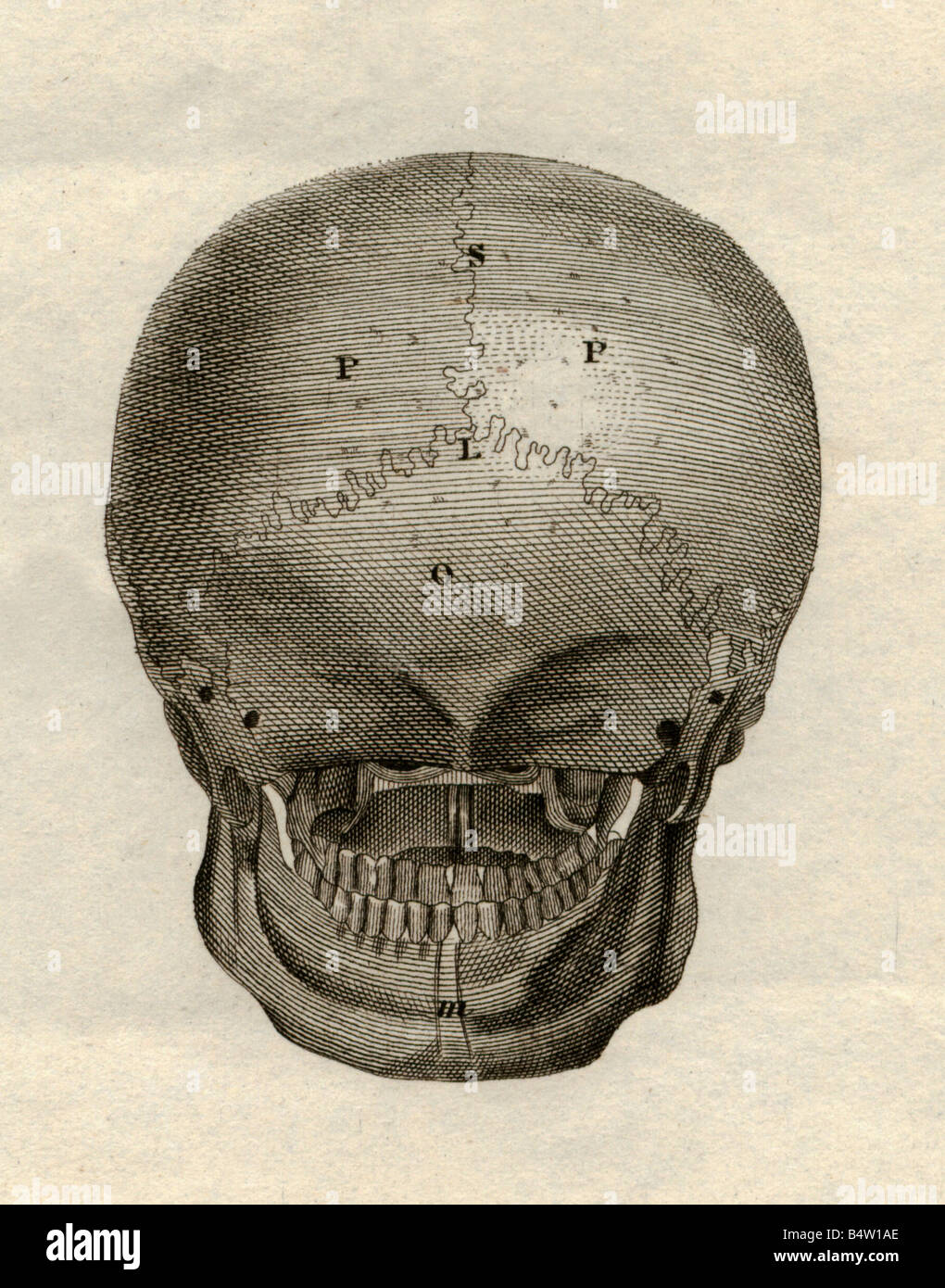
Cranium) is the skeleton of the head composed of 22 separate bones joined together primarily by sutures. First, the lambdoid suture connects the occipital bone to both parietal bones. The brain is connected with other anatomical structures by the nerves and blood vessels going through many foramina, and the largest foramen of the skull called the foramen magnum. The greater portion of the anterior floor is convex and grooved by the frontal lobe gyri. The occipital bone surrounds a large opening known as the foramen magnum.
Medicine Anatomy Skeleton Bones Skull Back View Steel Stock Photo Alamy from c8.alamy.com Pain in the back of your head at the base of your skull can cause your head to hurt with dull nagging persistent pains. Massaging the sinus pressure points at the back of the head. Frontal, sphenoid, ethmoid, occipital, parietal and temporal. It is also known as the calvarium. Overview, anterior skull base, middle skull base march 18, 2017. Bone back of head bigger on one side, bone back of the head, bone on back of skull, bone spur back of skull, skull bone back of head, bone, bone back of head bigger. The occipital bone houses the back part of the brain and is one of seven bones that come together to form the skull. The skull is composed of two parts:
In the adult, the skull consists of 22 individual bones, 21 of which are immobile and united into a single unit.
ads/bitcoin2.txt
The occipital bone is the only bone in your head that connects with your cervical spine (neck). Skull, skeletal framework of the head of vertebrates, composed of bones or cartilage, which form a unit that protects the brain and some sense organs. It also covers some common conditions and injuries that can affect the back. The skull is a strong, bony capsule that rests on the neck and encloses the brain. 1 also, some people are more prone to headaches than others. This portion of the skull base consists of the orbital portion of the frontal bone. There are four pairs of sinuses, named for the bones that they're located in: The skull base offers support from the bottom of the brain. Frontal, sphenoid, ethmoid, occipital, parietal and temporal. It is formed by a chain of 33 interconnected vertebrae and their intervening joints. Conditions associated with encephaloceles include hydrocephalus (excess accumulation of cerebrospinal fluid in the brain), developmental delays, microcephaly (an abnormally small head), paralysis and seizures. Skull system anatomy of skull anatomy skull skull lateral view skull anatomy occipital medical skeleton named facial bone anatomy mandible maxilla human anatomy lateral skull anatomy. The brain is connected with other anatomical structures by the nerves and blood vessels going through many foramina, and the largest foramen of the skull called the foramen magnum.
ads/bitcoin3.txt
ads/bitcoin4.txt
ads/bitcoin5.txt
0 Response to "Back Of Skull Anatomy : Skull Notes"


0 Response to "Back Of Skull Anatomy : Skull Notes"
Post a Comment