Upper Back Anatomy / Back Muscles Anatomy Of Upper Middle Lower Back Pain In Diagrams Goodpath
Upper Back Anatomy / Back Muscles Anatomy Of Upper Middle Lower Back Pain In Diagrams Goodpath. Back anatomy the back is the body region between the neck and the gluteal regions. License image the deltoid, teres major, teres minor, infraspinatus, supraspinatus (not shown) and subscapularis muscles (not shown) all extend from the scapula to the humerus and act on the shoulder joint. Your thoracic spine gives your upper body strength and stability and helps you lift heavy objects. Muscle anatomy app 12 photos of the muscle anatomy app best muscle anatomy app, best muscle anatomy app for android, free muscle anatomy app, human muscle anatomy app, muscle anatomy app ipad, human muscles, best muscle anatomy app, best muscle anatomy app for android, free muscle anatomy app, human muscle anatomy app, muscle. Both the deltoid and the trapezius are firmly attached to …
ads/bitcoin1.txt
The upper back muscles of the rhomboids and the trapezius are responsible for many of the movements of the scapula which in turn plays a huge role in the stability and mobility of the shoulder. Muscle anatomy app 12 photos of the muscle anatomy app best muscle anatomy app, best muscle anatomy app for android, free muscle anatomy app, human muscle anatomy app, muscle anatomy app ipad, human muscles, best muscle anatomy app, best muscle anatomy app for android, free muscle anatomy app, human muscle anatomy app, muscle. The extrinsic back muscles, which lie most superficially on the back. See upper back stock video clips. In the upper back region, the trapezius, rhomboid major, and levator scapulae muscles anchor the scapula and clavicle to the spines of several vertebrae and the occipital bone of the skull.
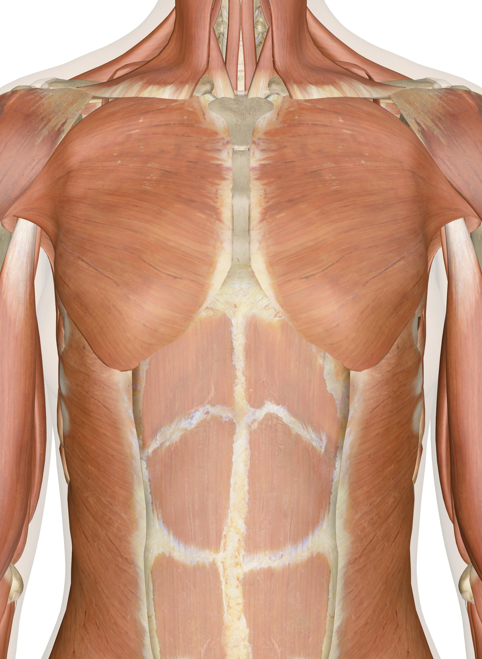
Muscles Of The Chest And Upper Back from innerbody.imgix.net Powerful muscles that move the head and arms attach to these bones as well. Upper right back pain under shoulder blade. In front of, front posterior: This muscle is located on the upper portion of the back anatomy, underneath the trapezius. The top of the cervical spine connects to the skull, and the bottom connects to the upper back at about shoulder level. In the upper back region, the trapezius, rhomboid major, and levator scapulae muscles anchor the scapula and clavicle to the spines of several vertebrae and the occipital bone of the skull. Anatomy of the upper back and middle back (thoracic spine) your upper and middle back starts from just below your neck and extends to about 5 inches below your shoulder blades. The superficial and intermediate muscles do not develop in the back, and are classified as extrinsic muscles.
The complexity of this region means that dysfunction can occur either due to injury or progressive pain and degeneration.
ads/bitcoin2.txt
Back anatomy the back is the body region between the neck and the gluteal regions. The upper portion is that trapezoid shape visible from the front of the body. These include the cervical vertebrae in the neck, the thoracic vertebrae of the ribcage in the upper and middle back, the lumbar vertebrae in the lower back, and the vertebrae that are part of the pelvis. An anatomy lesson is a good place to start. The bones of the chest and upper back combine to form the strong, protective rib cage around the vital thoracic organs such as the heart and lungs. Near, closer to the origin dorsal: Muscle strain, sprains, and spasms can affect the rhomboid muscles, which are located in the middle of the shoulder blades.this pain is mostly felt in. See upper back stock video clips. It originates from the base of the skull, along the nuchal ligament and the 7th cervical vertebra , which is that bony landmark on the back of your neck. Anatomy of the upper back and middle back (thoracic spine) your upper and middle back starts from just below your neck and extends to about 5 inches below your shoulder blades. This article will help you understand key anatomical structures in the skull and spine, with the goal of helping you better understand your condition. The complexity of this region means that dysfunction can occur either due to injury or progressive pain and degeneration. After, behind, following, toward the rear distal:
Muscle anatomy app 12 photos of the muscle anatomy app best muscle anatomy app, best muscle anatomy app for android, free muscle anatomy app, human muscle anatomy app, muscle anatomy app ipad, human muscles, best muscle anatomy app, best muscle anatomy app for android, free muscle anatomy app, human muscle anatomy app, muscle. They originate from the vertebrae and insert into the scapulae. It comprises the vertebral column (spine) and two compartments of back muscles; These muscles are divided into superficial and intermediate. The back muscles are divided into two large groups:
Anatomy Of The Upper Back from www.spineuniverse.com Powerful muscles that move the head and arms attach to these bones as well. It comprises the vertebral column (spine) and two compartments of back muscles; After, behind, following, toward the rear distal: In front of, front posterior: Upper back pain rear view of spine back pain spine sports spine spine surgery spine white background back ache x human anatomy illustration human anatomy on white background upper body stretch. An anatomy lesson is a good place to start. Related posts of upper back muscle diagram muscle anatomy app. The iliocostalis muscles are furthest from the spine.
Both the deltoid and the trapezius are firmly attached to …
ads/bitcoin2.txt
As a result of overuse or strenuous activity, at times these tendons tend to get inflamed resulting in painful symptoms. The complexity of this region means that dysfunction can occur either due to injury or progressive pain and degeneration. The upper back originates at the base of your neck, incorporates both shoulders and extends down to mid spine, including your ribs. They originate from the vertebrae and insert into the scapulae. The main superficial muscles of the back are the following: The cervical spine is the top part of the spine. Upper back pain rear view of spine back pain spine sports spine spine surgery spine white background back ache x human anatomy illustration human anatomy on white background upper body stretch. Try the injurymap exercise app now. The back functions are many, such as to house and protect the spinal cord, hold the body and head upright, and adjust the movements of the upper and lower limbs. Back muscles anatomy here include the trapezius, latissimus dorsi, rhomboid and levator scapulae. If you've been diagnosed with an upper cervical (neck) disorder, it's important to learn as much as you can about it. It runs from the neck to the upper back. Looking for a solution to your back pain problem?
Powerful muscles that move the head and arms attach to these bones as well. It is very stiff, and the thoracic spine has a limited range of motion. License image the deltoid, teres major, teres minor, infraspinatus, supraspinatus (not shown) and subscapularis muscles (not shown) all extend from the scapula to the humerus and act on the shoulder joint. The cervical spine is the top part of the spine. Back anatomy the back is the body region between the neck and the gluteal regions.
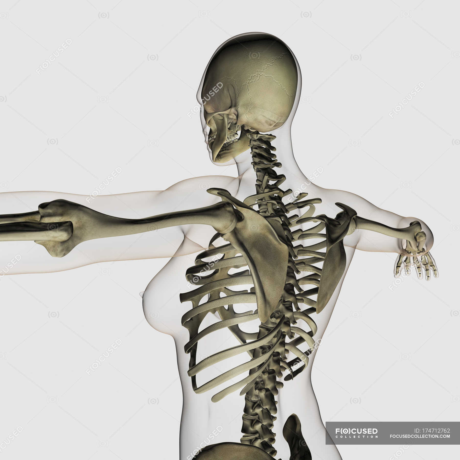
Three Dimensional View Of Female Upper Back And Skeletal System Anatomy Healthcare Stock Photo 174712762 from st.focusedcollection.com The cause may be poor posture (such as forward head posture) or any type of irritation of the large back and shoulder muscles, including muscle strain or spasms. Related posts of upper back muscle diagram muscle anatomy app. Upper right back pain under shoulder blade. License image the deltoid, teres major, teres minor, infraspinatus, supraspinatus (not shown) and subscapularis muscles (not shown) all extend from the scapula to the humerus and act on the shoulder joint. This article will help you understand key anatomical structures in the skull and spine, with the goal of helping you better understand your condition. Back anatomy the back is the body region between the neck and the gluteal regions. Near, closer to the origin dorsal: The deep muscles develop embryologically in the back, and are thus described as intrinsic muscles.
The cervical spine protects the nerves connecting to.
ads/bitcoin2.txt
After, behind, following, toward the rear distal: The cervical spine protects the nerves connecting to. Try the injurymap exercise app now. The back functions are many, such as to house and protect the spinal cord, hold the body and head upright, and adjust the movements of the upper and lower limbs. As a result of overuse or strenuous activity, at times these tendons tend to get inflamed resulting in painful symptoms. The cause may be poor posture (such as forward head posture) or any type of irritation of the large back and shoulder muscles, including muscle strain or spasms. This article will help you understand key anatomical structures in the skull and spine, with the goal of helping you better understand your condition. Away from, farther from the origin proximal: The complexity of this region means that dysfunction can occur either due to injury or progressive pain and degeneration. This is a tutorial to quickly s. It is very stiff, and the thoracic spine has a limited range of motion. The back muscles are divided into two large groups: The extrinsic back muscles, which lie most superficially on the back.
ads/bitcoin3.txt
ads/bitcoin4.txt
ads/bitcoin5.txt
0 Response to "Upper Back Anatomy / Back Muscles Anatomy Of Upper Middle Lower Back Pain In Diagrams Goodpath"


0 Response to "Upper Back Anatomy / Back Muscles Anatomy Of Upper Middle Lower Back Pain In Diagrams Goodpath"
Post a Comment